
How to perform the Ocular Counter Roll (OCR) test
What is the ocular counter roll test?
The ocular counter roll test is a clinical test to assess otolith function, which takes advantage of a torsional eye tracking algorithm. The test is done with video recording, so it is referred to as a video Ocular Counter Roll (vOCR) test. When the head is tilted, the eyes react with a torsional counter roll. This is a called an ocular counter roll (OCR), which is initiated by the vestibulo-ocular reflex (VOR).
Calibration
To perform the test, you should first calibrate the Torsion eye tracker. This allows the algorithm to identify the iris and track the movements of the eyeball.
First, select the Ocular Counter Roll protocol from the test menu.

Figure 1 – Test menu
Then select the calibration option.
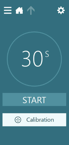
Figure 2 – Calibration selection
Once in the calibration menu, you can perform 2 tasks:
- Calibrate the Torsion tracker
- Calibrate the eyes to the TV monitor
First, calibrate the Torsion tracker. You can use the Auto Detect button to find the appropriate iris area for tracking. If the white tracking circles are not encompassing enough of the iris, you can use the manual slider adjust bars to make small adjustments.
![]()
Figure 3 – Calibrate torsion tracker
When you are satisfied that the tracker is stable around the eyes, then use the Set Reference button to finish the calibration.
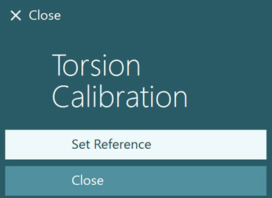
Figure 4 – Set torsion reference
Torsion calibration should be completed with the cover off, if performing the OCR test with the cover off. This quick guide overviews the process for testing with the cover off. If you elect to perform the OCR test with the cover on, torsion calibration should be completed with the cover on.
Next, calibrate the eyes to the TV monitor (if this has not been already completed for other oculomotor tests). Instruct the patient to sequentially look at each of the five dots. The test will automatically stop when the calibration is completed.

Figure 5 – Video eye calibration
Performing the test
Once the system is calibrated, you are ready to begin testing. The TV screen will have a solid background and a green center fixation dot. Have the patient look at the dot while with the head centered and hold there for a minimum of 10 seconds. Try to ensure the sensor is as close to 0 for accurate measurements. Press the enter key on the remote to mark the next section and then rotate their head and shoulders 30 degrees to the left.
When you are in the correct 30-degree position hold there for at least 30 seconds or until the eye tracing stabilizes. Press the enter key again and bring the patient back to the center. Try to ensure the sensor is as close to 0 for accurate measurements. Hold there another minimum of 10 seconds. Press the enter key again and rotate the head and shoulders 30 degrees to the right and again hold there for at least 30 seconds or until the eye tracing stabilizes. This is called a static ocular counter roll test because the patient’s head is held still in each position.

Figure 6 – Positioning the patient
If you have a VORTEQ Sensor, then you can use the 3D head model to guide you in properly positioning the head and shoulders to the proper angle. The default angle is 30 degrees. The green target bar will guide you and the patient in the position. Exact degree will also be available for accuracy.
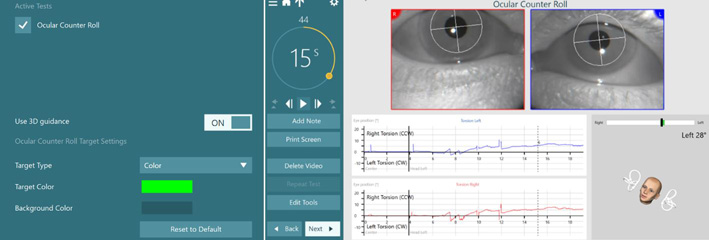
Figure 7 – VORTEQ sensor and 3D head model guidance
Results
In the results section, you will see the eye position (in degrees) for left and right head tilts.

Figure 8 – Graphical display of ocular counter roll test results
You can measure the degree of torsion using the edit functions to add in numerical values. The ocular counter roll slow phase movement is plotted. When the head is tilted to the left shoulder, you will see a right slow torsion counterclockwise (to the examiner) eye movement. When the head is tilted to the right shoulder, you will see a slow torsion left in the clockwise direction.
In the static ocular counter roll test, we are only interested in the final position of the eye. This is represented as the flat area where the eye is still (static) after the head is held in position for at least 10 seconds. The dynamic eye movements during the head movements are not analyzed.
For further assistance, if needed, please refer to the Instructions for Use and Additional Information manuals.
Presenter
