
Understanding torsional eye movements
Description
Measurement and analysis of torsional eye movements are available in the VisualEyes™ software. These measurements provide an assessment of the vertical semicircular canals, otoliths, and their central pathways during the Advanced Dix-Hallpike and Ocular Counter Roll (OCR) tests.
Quantification of torsional eye movements adds an important tool to the evaluation of dizzy patients. However, deciphering the tracings and understanding the direction of torsional eye movements can be confusing to many users. Part of this confusion is related to the novelty of these recordings. The other part of the confusion is related to the historically contradictory and unclear terminologies used to describe torsional eye movements in the literature.
This paper provides a brief review of the torsional eye movements and suggests logical terminologies that can be used to describe them. Some of the more complicated topics associated with torsional eye movements, such as the Listing’s plane, will not be discussed here.
Related course: Advances in VNG
Review of horizontal and vertical eye movements
The eyes can move in three dimensions:
- Yaw (horizontal)
- Pitch (vertical)
- Roll (torsion)
For horizontal and vertical eye movements, the center of rotation is located approximately 13‑15 mm behind the cornea (Figure 1A&B).

There is a slight difference between the horizontal and vertical centers of rotation, but we will not consider that for the purposes of this article. The reference line for horizontal and vertical eye movements (zero axis represented by the dashed line) is the line that connects the center of rotation to the center of pupil when the eye is looking straight ahead. When the eye moves horizontally, the angle of the line that connects the center of rotation to the new pupil center is used to measure the horizontal eye movement (Figure 1A bottom graph).
By convention, the angle is assigned positive numbers when the eye moves to the right of the subject from the reference line. Conversely, the angle is assigned negative numbers when the eye moves to the left of the subject from the reference line. Therefore, rightward eye movements are represented by tracings that increase in value whereas leftward eye movements are represented by tracings that decrease in value.
Figure 2 illustrates the horizontal eye movements during the gaze test.
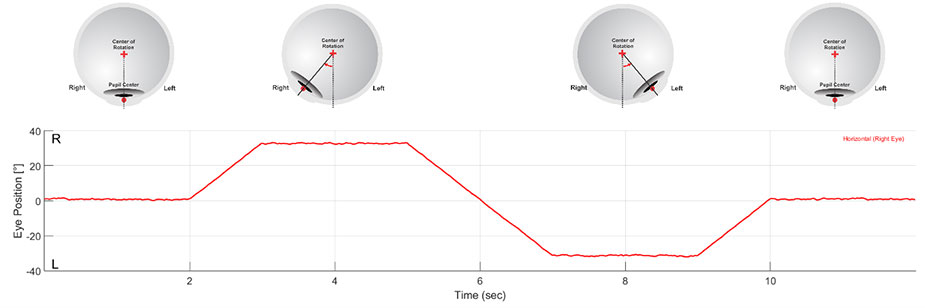
The tracing is at 0° for the first 2 seconds signifying that the center of the pupil is at the reference point and the eye is looking straight ahead. The eye angle increases signifying that the center of the pupil is moving rightward.
The eye remains at the right gaze until t=4 seconds. From there, the eye angle decreases signifying that the center of the pupil is moving leftward. After remaining in the left gaze position, the eye angle increases again signifying that the center of the pupil is moving rightward. The tracing returns to 0° when the eye is at the center gaze again.
The key here is that the definitions for right and left directions are made in reference to the subject.
The convention for vertical eye movements (Figure 1B) is very similar with upward eye movements represented by tracings that increase in value and downward eye movements are represented by tracings that decrease in value.
The conventions for horizontal and vertical eye movements are well-understood by those who have experience with eye movement recordings. Yet, novice users may still be unclear when those conventions are used to describe nystagmus.
By definition, nystagmus consists of a slow movement in one direction followed by a fast movement in the opposite direction. Therefore, it is meaningless to assign a direction to nystagmus without specifying which component is being described. This issue is addressed by adding the term “beat” to the direction of nystagmus to signify the fast component of nystagmus.
For example, Figure 3A shows several seconds of left-beating nystagmus (rightward slow phase) followed by several seconds of right-beating nystagmus (leftward slow phase).
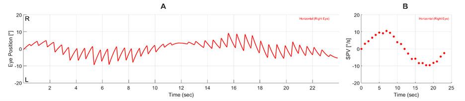
The direction of fast phases indicates either excitation of the ipsilateral lateral canal pathways or inhibition (lesion) of the contralateral lateral canal pathways. The intensity of nystagmus is measured by the slow-phase velocity (SPV). Positive numbers represent rightward slow phase or left-beating nystagmus, and negative numbers represent leftward slow phase or right-beating nystagmus (Figure 3B).
Torsional eye movements
When analyzing torsional eye movements, it is best to use conventions that are similar to those used for horizontal eye movements and to preserve the relationship between the direction of nystagmus and the structures that produce them.
For torsional eye movements, the center of rotation is the center of pupil, and the reference line (zero axis) is the line that connects the upper pole of the eye to the center of the pupil when the eye is looking straight ahead (Figure 1C). When the eye rotates in the roll plane, the angle of the line that connects the center of pupil to the new location of the upper pole can be used to measure the torsional eye movement.
To preserve the convention used for horizontal eye movements, we can consider the angle of the eye following movement of the upper pole to the right of the subject as right torsion and assign positive numbers to it. Conversely, left torsion occurs when the upper pole of the eye torsions to the left of the subject. Negative numbers are assigned for left torsion.
As such, rightward torsional eye movements will be represented by tracings that increase in value whereas leftward torsional eye movements are represented by tracings that decrease in value.
To differentiate between horizontal and torsional eye movements, one should always add the word torsion when describing the direction of torsional eye movements, for example, rightward torsion.
Figure 4 shows torsional eye movements that are similar to the horizontal eye movements in Figure 2.
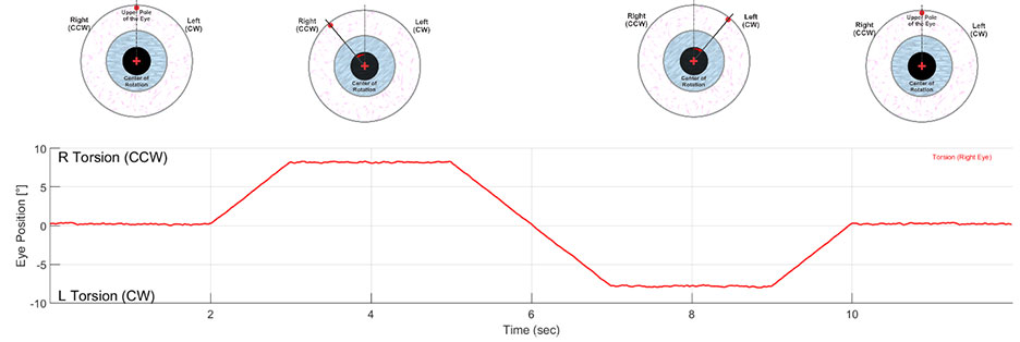
The tracing is at 0° for the first 2 seconds signifying that the upper pole of the eye is at the reference point and the eye is looking straight ahead. After that, the torsional angle increases signifying that the upper pole of the eye is rotating toward the subject’s right. The eye remains at the position until t=4 seconds.
From there, the torsional angle decreases signifying that the upper pole of the eye is rotating toward the subject’s left. After remaining in this position, the torsional angle increases again signifying that the upper pole of the eye is rotating rightward. The tracing returns to 0° when the upper pole of the eye is again at the reference point.
Using the above convention for torsional eye movements retains the direction reference to the subject and is consistent with the convention used for horizontal movements without adding new terminologies. Unfortunately, torsional eye movements are often described in the literature using the terms clockwise (CW) and counterclockwise (CCW). These terms describe the movement of any point on the iris from the examiner’s point of view. As such, CW torsion will be equivalent to left torsion and CCW torsion will be equivalent to right torsion (Figure 4).
The appeal of this terminology is that the examiner can describe the movement he/she sees without referencing the subject. However, this terminology may make it more difficult in identifying the side of lesion or the side of excitation that is generating torsional eye movement responses.
Another shortcoming is that the terms CW and CCW often imply motion. That is fine for describing nystagmus following tests such as the Dix-Hallpike maneuver but may cause ambiguity for tests such as the ocular counter-rolling (OCR) test, where the head tilts cause static torsion.
Some clinicians have tried to use the terms CW and CCW from the subject’s point of view. This attempt does not address the fundamental issue in identifying the side of excitation or inhibition in a manner that is consistent with horizontal eye movements. Furthermore, it nullifies the main reason for using CW and CCW terminologies, which is for the examiner to describe the torsion without referencing the subject. Instead, the examiner must imagine looking from behind the subject’s eyes and describing the direction of torsion. That does not seem to be an easy task for Dix-Hallpike and similar maneuvers.
Regardless of which terminologies are used, the description of nystagmus directions should follow the same convention for vertical and horizontal nystagmus. For example, for nystagmus where the upper pole of the eye undergoes fast movements to the right can be called right-beating torsional nystagmus. This can also be called CCW-beating nystagmus. To reference the slow phase of the same nystagmus, one can use the term leftward slow torsion or CW slow torsion.
In vestibular testing, torsional eye movements are usually generated by head movements, which often cause both dynamic and static torsion. Figure 5A shows a tracing generated during the OCR test and describes the direction of torsional eye movements during different parts of the test.
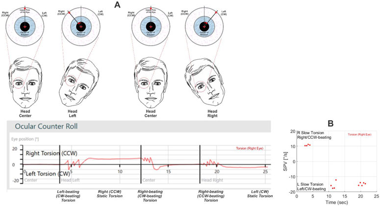
First, the head is in the upright position with the eye looking straight ahead. Accordingly, the tracing shows 0° of torsion. Then the head is rotated to the left in the roll plane. Initially, there is a dynamic response consisting of a few beats of left-beating torsional nystagmus. This is similar to when the head is rotated left in the horizontal plane, which generates left-beating horizontal nystagmus.
One can use the term CW-beating nystagmus here but the similarity with the horizontal responses is no longer present. After the dynamic component subsides, there is a rightward (CCW) static torsion. When the head is returned to the upright position, a few beats of right-beating (CCW-beating) nystagmus can be seen followed by the upper poll of the eye returning to the reference point.
When the head is tilted to the right, a few beats of right-beating (CCW-beating) torsional nystagmus are seen followed by a static leftward (CW) torsion. Again, describing the torsion based on the subject’s point of view preserves the correspondence to the horizontal nystagmus and implies the same pattern of excitation or inhibition for the involved labyrinthine structures.
Summary
There are different ways to describe torsional eye movements. The preferred convention is to be consistent with the convention used for horizontal and vertical eye movements and describe the torsion of the upper pole of the eye with respect to the subject. The terms CW and CCW, if one chooses to use them, should be used from the examiner’s point of view. In that case, CW torsion will be equivalent to left torsion and CCW torsion will be equivalent to right torsion.
About the authors
Kamran Barin, Ph.D., is Assistant Professor Emeritus, Department of Otolaryngology-Head & Neck Surgery and Department of Speech & Hearing Science, The Ohio State University. He established and served as the Director of Balance Disorders Clinic at the Ohio State University Medical Center for over 25 years until his retirement in June 2011. He received his Master’s and Doctorate degrees in Electrical/Biomedical Engineering from the Ohio State University. He has published over 80 articles and book chapters and has taught national and international courses and seminars in different areas of vestibular assessment and rehabilitation.
Michelle Petrak, Ph.D., is the Director of Clinical Audiology and Vestibular Research for Interacoustics. She is located in Chicago where she is a licensed private practice clinical audiologist at Northwest Speech and Hearing (NWSPH). Dr. Petrak received her doctorates in Electrophysiology (1992) and Biomolecular Electronics (1994) from Wayne State University and her Masters in Audiology in 1989.