
Dynamic Visual Acuity (DVA)
What is the Dynamic Visual Acuity (DVA) test?
Dynamic Visual Acuity (DVA) is a behavioral assessment of the Vestibulo-Ocular Reflex (VOR) in response to head movements. During the test, patients move their heads at a consistent speed, while an optotype decreases in size until the software detects the DVA threshold. This assessment is often paired together with the Gaze Stabilization Test (GST).
If you have purchased the VORTEQ™ Assessment bundle, you will be able to perform the DVA test.

Figure 1 - Tests in the VORTEQ™ Assessment bundle
Screen setup
Select the Optotype Stimuli display source and set the screen size and patient distance in the System Default Settings before beginning the first test. A suggested patient distance will be identified for your screen dimensions. If the distance is too much for your room set up, then choose a smaller display input.

Figure 2 - Optotype stimuli settings for DVA
Protocol setup
The default protocol is automatic, but you can also choose a manual target presentation. You can choose the options in Summary Parameters.

Figure 3 - Summary Parameters protocol setup for DVA
The default starting head speed is 100 degrees per second, but you can adjust that if needed.

Figure 4 - Test Parameters protocol setup for DVA
The metronome should be “ON” to give the patient feedback on how fast to move their head. The metronome sound will stop after each optotype appears to give the patient time to enter their response with the remote control. The sound will start again when they move their head.
Preparing for the test
To begin testing, select Dynamic Visual Acuity from your drop-down test menu.

Figure 5 - Test menu for DVA
A reminder will pop up to make sure you remove the VORTEQ™ IMU from the goggles and attach it to the headband.
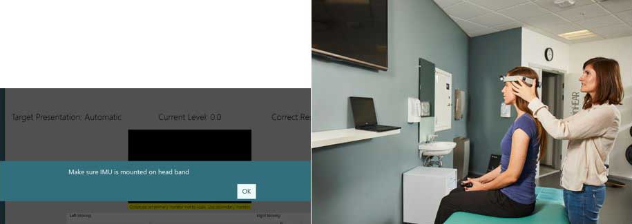
Figure 6 (a) Reminder to put the VORTEQ IMU on the headband and (b) Patient wearing DVA headband during DVA test
If the IMU is not turned on, you will see this error message.
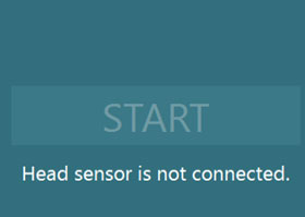
Figure 7 - Start button is inactive if the sensor is not turned on
Once you have mounted the headband on the patient’s head and turned on the sensor, you can hand the patient the remote control. Instruct the patient to press the arrow that matches the direction of the optotype they see during the testing.

Figure 8 - Remote control for DVA test
If they do not know the direction, they can tell you “I don’t know” and then you can click on the “?” on the screen to enter the “I don’t know” response for them.
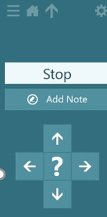
Figure 9 - Patient options for optotype direction
How to perform the Static Visual Acuity (SVA) test
If you have already performed the Static Visual Acuity (SVA) test (in GST test), you can copy the results over using the “Copy Static Result” button.
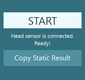
Figure 10 - Copy Static Result
If you have not performed the SVA test yet, then you must complete it before moving on.
For the SVA test, the patient keeps their head still and responds to the direction of the optotype that appears in the white square on the TV screen.
You will see this test screen.

Figure 11 SVA test - clinician’s screen
When you press start, you will see the optotype direction that the patient sees and the button that they are pressing.
SVA results
When you have completed the test, you will see the patient’s static acuity score.

Figure 12 - SVA results
How to perform the DVA test
Have the patient move their head so that the speed of their head movement peaks in the green area of the velocity bar. Their head movement is the solid line. The acceptable range for head movement is the green shaded area. When their head movement peaks in this area, the optotype will appear. You can see this on the screen as the direction arrows will become highlighted in white when the optotype has appeared.

Figure 13 - Proper head speed
If the patient’s head movement is too slow, then you will see a red solid line that peaks before the green shaded area. You will see the progress of the head movement as a grey bar that ends at the red solid line. Instruct the patient to move their head faster and follow the beat of the metronome so they can reach the green shaded area.

Figure 14 - Head speed too slow
If the patient’s head movement is too quick, then you’ll see a red solid line that peaks after the green shaded area. You will see the progress of the head movement as a red shaded area that ends at the red solid line. Instruct the patient to slow down their head movements so they can reach the green shaded area.

Figure 15 - Head speed too fast
DVA results
After you complete all four runs (left, right, up, down), you will see a summary graph of the patient’s dynamic visual acuity for both horizontal and vertical eye movements.

Figure 16 - Completed test summary screen
There are no default thresholds, but you can choose to add your own suggested thresholds. If you provide your own thresholds, then any data points falling in the grey shaded areas would be outside of the suggested normal threshold ranges. This would show that the patient has trouble staying focused on the target at this head speed.
For further help, please refer to the Instructions for Use and Additional Information manuals.
Presenter
