
How to test otolith function
For many years, we have used the term ‘vestibular function testing’ with the implication that we are testing the entire vestibular system.
In fact, most of our tests, such as the caloric test, vHIT, rotary chair, and even the Dix-Hallpike test, are tests of semicircular canal function and do not test the otoliths.
In the 1990s, with the introduction of vestibular-evoked myogenic potentials (VEMPs), we finally had a quantitative test for the otoliths.
Although cervical VEMP (cVEMP) and ocular VEMP (oVEMP) are important additions to the vestibular test battery, they need a different hardware/software environment.
It would be nice to be able to test the otoliths within the same environment that we use for the more traditional tests such as the caloric or the rotary chair test.
VisualEyes™ offers two options for this purpose:
- Ocular counter roll (OCR)
- Dynamic subjective visual vertical (SVV)
I will address each below, and refer to the video just below throughout the article, using time stamps to make sure you only watch the part that is relevant for each section.
1. Ocular counter roll (OCR)
The first option is the ocular counter roll (OCR) test.
It consists of tilting the head toward either the right or left shoulder in the roll plane.
As you can see in this portion of the video, the patient’s head starts upright and then the clinician tilts it left and holds it there for several seconds.
Then the clinician brings the patient’s head back to the upright position and tilts it to the right.
Three-dimensional eye movements are recorded throughout.
The head tilt provokes the otolith-ocular reflex, which causes compensatory eye movements by generating static torsion of the eyes in the opposite direction.
The movement also generates vertical skew deviation.
In a person with normal otolith function, there is no static torsion in the upright head position and the responses for the right and left head tilts are symmetrical (Figure 1).
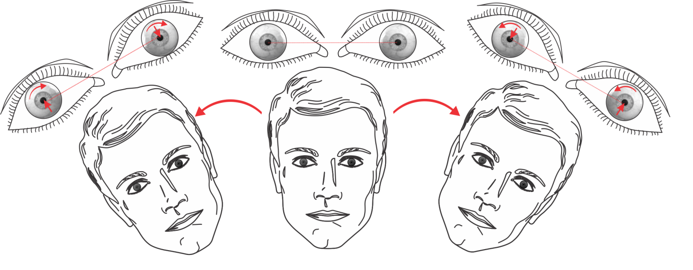
Figure 1: Eye movements in response to head tilts in the roll plane.
OCR components
In this portion of the video, you can see the torsional eye movement recordings during the ocular counter roll test.
First the head is upright and after a few seconds, the head is tilted to the left.
Again after a few seconds, the head is brought back to the upright position and tilted to the right.
The response consists of two components:
- The dynamic component
- The static component
Dynamic OCR component
The dynamic component is the transient nystagmus that begins during or shortly after the head tilt and lasts for several seconds.
This component is meditated primarily by the vertical semicircular canals with minimal contribution from the otoliths.
Static OCR component
The static component is the persistent torsion of the eye position that lasts for the duration of head tilt.
This component is mediated by the otoliths and their central pathways.
Unilateral otolith lesion
In the head upright position, a patient with a unilateral otolith lesion will have static torsion of the eyes toward the side of lesion and skew deviation as if the head was tilted contralateral to the side of lesion.
Also, the ocular tilt reaction (OTR) becomes asymmetrical with smaller responses for head tilts toward the side of lesion.
In this portion of the video, you can see the ocular counter roll test results in a patient with otolith dysfunction.
The oVEMPs show smaller responses for the right side compared to the left side.
The OCR responses for the left head tilt are larger than those for the right head tilt.
This asymmetry is consistent with the oVEMP results and signifies a right utricular abnormality.
Key takeaway: The main clinical utility of the OCR test is that it provides a quantitative method to document otolith abnormalities.
2. Dynamic subjective visual vertical (SVV)
The second option in VisualEyes™ that allows us to test otolith function is the capability to measure the dynamic subjective visual vertical (SVV).
SVV is the perceptual equivalent of the ocular counter roll.
You are probably familiar with static SVV, which you can measure with something as simple as the ‘bucket’ test or with more sophisticated goggles.
But, as mentioned above, it is also possible to measure dynamic SVV.
How to measure dynamic SVV
To measure dynamic SVV, you need a rotary chair that is capable of off-axis (eccentric) rotation.
During on-axis rotation, responses from the right and left utricles cancel each other out.
If we move the chair such that the axis of rotation goes through one of the utricles, then the off-axis rotation generates centrifugal or centripetal forces and stimulates the contralateral utricle.
After a prolonged rotation at constant velocity, the canal responses subside and the utricular response consisting of ocular counter-rolling becomes clear.
This can be measured by the SVV test during eccentric rotation.
SVV test with eccentric rotation (step-by-step)
As you can see in this portion of the video, a line is projected on the enclosure wall and the subject can control its orientation with a remote.
This is shown with the chair stationary to get the static SVV.
But you can perform the measurement during eccentric rotation to get the dynamic SVV, which can identify dynamic otolith abnormalities.
Steps:
- Before the test begins, the chair is moved to an off-axis position. When moved to the left, you are testing right utricular function, and vice versa.
- Then the chair is slowly accelerated toward the final velocity of around 300 deg/sec.
- The patient is rotated at this peak velocity until the canal responses completely subside. At that point, the responses are mediated primarily by utricular function.
- After this prolonged rotation, the luminous red line is presented to the patient, and the patient can change the orientation of the line with a remote until it appears to be vertical.
Summary
VisualEyes™ offers two methods for evaluating otolith function within the same software environment used for videonystagmography (VNG) and rotary chair testing.
These are the ocular counter roll test and dynamic SVV during off-axis rotation, which provide a more complete evaluation of the vestibular system.
