Subscribe to the Interacoustics Academy newsletter for updates and priority access to online events
Training in VEMP
-
Why Use the CE-Chirp® in VEMP Testing?
-
VEMP Tuning: Clinical Application (1/3)
-
VEMP Tuning: Clinical Application (2/3)
-
VEMP Tuning: Clinical Application (3/3)
-
oVEMP: An Introduction
-
cVEMP: An Introduction
-
Course: Balance Testing for Beginners
-
Course: Balance Testing for Intermediates
-
How Many Sweeps in cVEMP Testing?
-
How to Diagnose SSCD with cVEMPs
-
VEMP and vHIT in Vestibular Neuritis Patients
-
Can You Use VEMPs to Diagnose Meniere's Disease?
-
Getting started: VEMP
-
Vestibular Evoked Myogenic Potentials - Introduction
-
Cervical VEMP - Patient Preparation for Assessment
-
Cervical VEMP - Protocol & Parameter Selection
-
Cervical VEMP - Running the Test
-
Cervical VEMP - Modifications of the Assessment
-
Ocular VEMP - Patient Preparation for Assessment
-
Ocular VEMP - Modifications of the Assessment
-
Ocular VEMP - Running the Test
-
Ocular VEMP - Protocol & Parameter Selection
-
Vestibular Evoked Myogenic Potentials (VEMP): A Complete Guide
-
oVEMPs: Una introducción
-
Introducción a los cVEMP
-
Evaluación otolítica: Más allá de los VEMPs
-
Potentiels myogéniques évoqués vestibulaires (VEMP)
-
Potenciales Miogénicos Evocados Vestibulares (VEMP): Una Guía Completa
Accounting for Muscle Asymmetries in cVEMP
Description
The Interacoustics Eclipse has several features to assist you with this technical challenge related to the cVEMP.
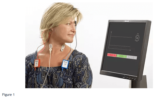
Firstly the instrument is equipped with an “EMG monitor” – in other words the system measures the myogenic activity associated with muscle contraction and provides visual feedback via a computer monitor so that the patient can maintain a target level of contraction. To avoid asymmetries of course the same target is used when testing the left and right cVEMP. The below figure (Figure 1) shows an example of this. Here the patient is having the right cVEMP measured, hence is gazing over the left shoulder to contract the right sternocleidomastoid muscle. The visual feedback consists of the scroll bar (at the bottom), which is colour coded and hovers in the green zone when the correct muscle tone is obtained. If either too much or too little muscle tone occurs (which can happen momentarily in some patients) then the scroll bar will go up or down into red zones accordingly, and the system will pause the averaging of these trials until the correct muscle tone is again obtained.
In addition to the scroll bar there is a dial (top right of screen) indicating test progress (% of requisite number of accepted sweeps) and this can help guide the patient as to how much longer they need to keep up their efforts. The top left shows a chart which plots the EMG amplitude (y-axis) over time. A consistent muscle contraction should produce a horizontal line.

After cVEMPs for each ear are collected the clinician can compare the EMG graphs to “eyeball” these charts and ensure there is a similar mean EMG, and overlapping variance. For example the below figure (Figure 2) shows the result after completion of right ear measures.

The visual feedback to the patient provides a way to help prevent muscle asymmetries from occurring. However, if asymmetries do occur then the Interacoustics Eclipse features a further tool to account for any muscle asymmetries and this provides a way to ensure valid interaural comparisons of saccular function despite muscle asymmetries; the feature is known as EMG scaling.
The VEMP amplitude is scaled in proportion to the tonic EMG activity, which is calculated from the pre-stimulus period. So, low muscle contraction would produce a scaled up VEMP and high muscle contraction would produce a scaled down VEMP according to the following equation:
Averaged VEMP response amplitude (μV) divided by root mean square of pre-stimulation EMG activity (μV)
Presenter

Get priority access to training
Sign up to the Interacoustics Academy newsletter to be the first to hear about our latest updates and get priority access to our online events.
By signing up, I accept to receive newsletter e-mails from Interacoustics. I can withdraw my consent at any time by using the ‘unsubscribe’-function included in each e-mail.
Click here and read our privacy notice, if you want to know more about how we treat and protect your personal data.
